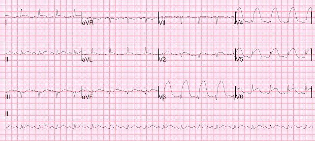
A middle-age male arrived by private car with chest pain. He immediately became unresponsive, before an ECG could be recorded.
He was resuscitated from ventricular fibrillation and this 12-lead was recorded:
Same ECG with annotation:
This ECG was recorded just before transportation to the cath lab:
A mid LAD 100% thrombotic occlusion was opened and stented. Door to balloon time was less than 60 minutes. Echo next day showed 57% EF. Peak troponin I was 250 ng/mL.
He was resuscitated from ventricular fibrillation and this 12-lead was recorded:
 |
| What are the wide complexes in the precordial leads? |
Same ECG with annotation:
This ECG was recorded just before transportation to the cath lab:
 |
| Sinus tachycardia at a rate of about 100. Massive ST elevation continues. |
A mid LAD 100% thrombotic occlusion was opened and stented. Door to balloon time was less than 60 minutes. Echo next day showed 57% EF. Peak troponin I was 250 ng/mL.


Hey Steve, it is extremely rare for a single PVC to terminate PSVT (especially if it is atrial tachycardia or AVNRT). You see if happen more often with AVRT (because the circuit involves the ventricle) but usually it just results in entrainment of the circuit for one beat and the tachycardia continues.
ReplyDeletethanks. very helpful!
DeleteSteve Smith
thank you for the interesting case. i was thinking svt + st elevation but when looking for signs of VT i noticed right axis deviation (positiv II/III leads). finding no other signs of vt this was still a bit misleading to me. also, in the follow up ecg after reperfusion axis deviation was "corrected" III now being negativ. any comments?
ReplyDeleteFirst ECG has an axis of about 90. Second is about 0. The second is clearly supraventricular. Is the first something else? I doubt it. Not sure why the axis changes.
DeleteThanks for the great comment.
Steve